The Lab
We are an experimental physics group in the Department of Physics at Oregon State University. We investigate applied physics questions related to carbon nanotube and graphene devices. The broader impact of our work ranges from energy conversion to molecular biology and medical diagnostics.
Several aspects of our work cross department boundaries. For example, in the field of photovoltaics we collaborate with materials scientists at OSU to manipulate and measure the properties of excitons in carbon nanotubes. In the field of nanobiosensors we work with molecular biologist Dr. Kerstin Blank to detect the electric fields around biogically active proteins and collaborate with researchers in the College of Pharmacy and the Department of Chemistry to identify low-concentration protein markers at the early stages of cancer.
Please contact Ethan Minot to learn more about opportunities for postdoctoral and PhD projects, and undergraduate research experience.
FundingWe are grateful for support by NSF, HFSP, ACS Petroleum Research Fund and ONAMI.
Lab facilities
The lab is equiped with an Asylum Research Atomic Force Microscope, a Fianium supercontinuum light source for scanning photocurrent spectroscopy, a carbon nanotube growth furnace (made by Kevek Innovations, a spin-off company from our research group), a graphene growth furnace for large-area graphene (up to 30 cm x 30 cm, Kevek Innovations), a probe station for electrical characterisation, fume hoods/wet benches for device fabrication and protein preparation.
Campus-shared facilities for photolithography and thin-film deposition systems (including direct write laser lithography and focussed ion beam lithography) are used to create new devices. Electron-beam lithography is available at University of Oregon (45 minute drive) and is currently being installed at the OSU Electron Microscopy Facility. Other nanofacilities are listed here.
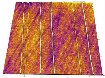
Figure 1 Atomic force microscope image (2 micron scan size) of single walled carbon nanotubes (~ 1 nm tall) grown on a quartz substrate. Van der Waals interactions between the CNTs and the quartz surface guide the growth direction.